Page 14 of 21
PA16.1-3 | Hemolytic Anemias — Practice Activities
Interactive Practice Activities
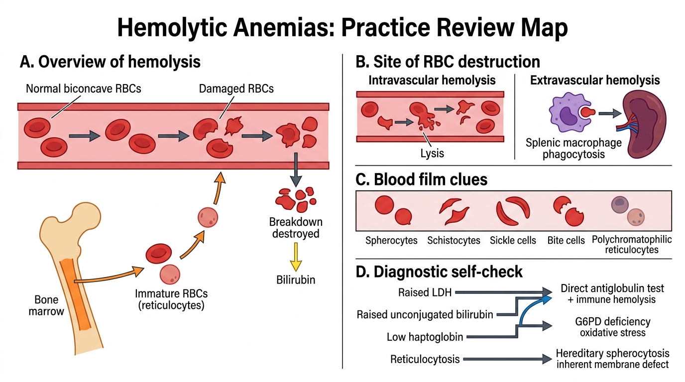
Hemolytic Anemias Practice Review Map
Panel A: Normal RBC, damaged RBC, bone marrow reticulocytosis, reticulocyte, hemoglobin breakdown, unconjugated bilirubin, anemia due to shortened RBC survival. Panel B: Intravascular hemolysis, free hemoglobin, hemoglobinuria, low haptoglobin, extravascular hemolysis, splenic macrophage, liver macrophage, splenomegaly. Panel C: Spherocyte, schistocyte, sickle cell, bite cell, polychromatophilic reticulocyte, peripheral blood smear. Panel D: Raised LDH, raised unconjugated bilirubin, low haptoglobin, reticulocytosis, direct antiglobulin test positive, immune hemolysis, G6PD deficiency, hereditary spherocytosis.
Reinforce the key terms and concepts from this module with these self-check activities.
Interactive practice: Crossword
Interactive practice: Flashcards
Interactive practice: Image Hotspots