Page 14 of 20
FM7.2 | Fall from Height & Vehicular Injuries — SDL Guide (Part 2)
Railway Injuries and Railway Spine
Railway injuries present a distinct forensic challenge because the mechanisms are multiple and the injuries are often extreme — the combination of high vehicle velocity, metal wheel-on-rail contact, and the victim's exposure to the rail environment produces injuries unlike other blunt force trauma. The forensic physician must distinguish between: injuries from the initial locomotive/carriage impact, injuries from the wheels passing over the body, and the specific condition of railway spine.
The initial locomotive impact may produce blunt force injuries similar to high-velocity vehicular impact — the victim is thrown, may be dragged by the undercarriage, and ultimately comes to rest with multiple injuries from several mechanisms. Wheel-over injuries are the most distinctive: when the train wheel passes over a limb or the body, it produces a characteristic wheel-mark injury — a patterned pressure necrosis and laceration corresponding to the wheel flange, which projects below the wheel's contact surface by approximately 1–2 cm. The flange produces a linear deep cut adjacent to a broad compression zone, distinguishing it from other crush injuries. The wheel's geometry (circumference, flange width) can be matched to injury dimensions.
Railway mutilation is extreme tissue destruction from the combination of wheel compression, tearing, and the rail-between-wheel mechanism — which can produce complete amputation, degloving, or bisection of the body. The forensic significance is in determining the ante-mortem vs post-mortem nature of the wounds (vital reaction, haemorrhage in wound margins) to distinguish death before derailment from death caused by the train.
Railway spine is a specific term in forensic medicine, described by Reddy's and Modi's, referring to a neurological injury of the spinal cord and/or nerve roots resulting from jarring, jolting, or sudden deceleration forces in railway accidents — without necessarily causing an overt vertebral fracture. The mechanism is functional or micro-structural disruption of neural tissue from the vibration and jolting forces transmitted through the spine during a railway accident. Railway spine presents as a spectrum from transient paresis (cord concussion) to permanent paraplegia (cord contusion or laceration at a single level) with preservation of the bony spinal column. The forensic significance is twofold: (1) a plaintiff claiming permanent spinal injury from a railway accident may have no fracture on plain X-ray — the physician must explain that neurological injury without fracture is a recognised entity in railway accidents; (2) distinguishing railway spine from malingering requires careful neurological documentation and serial examination.
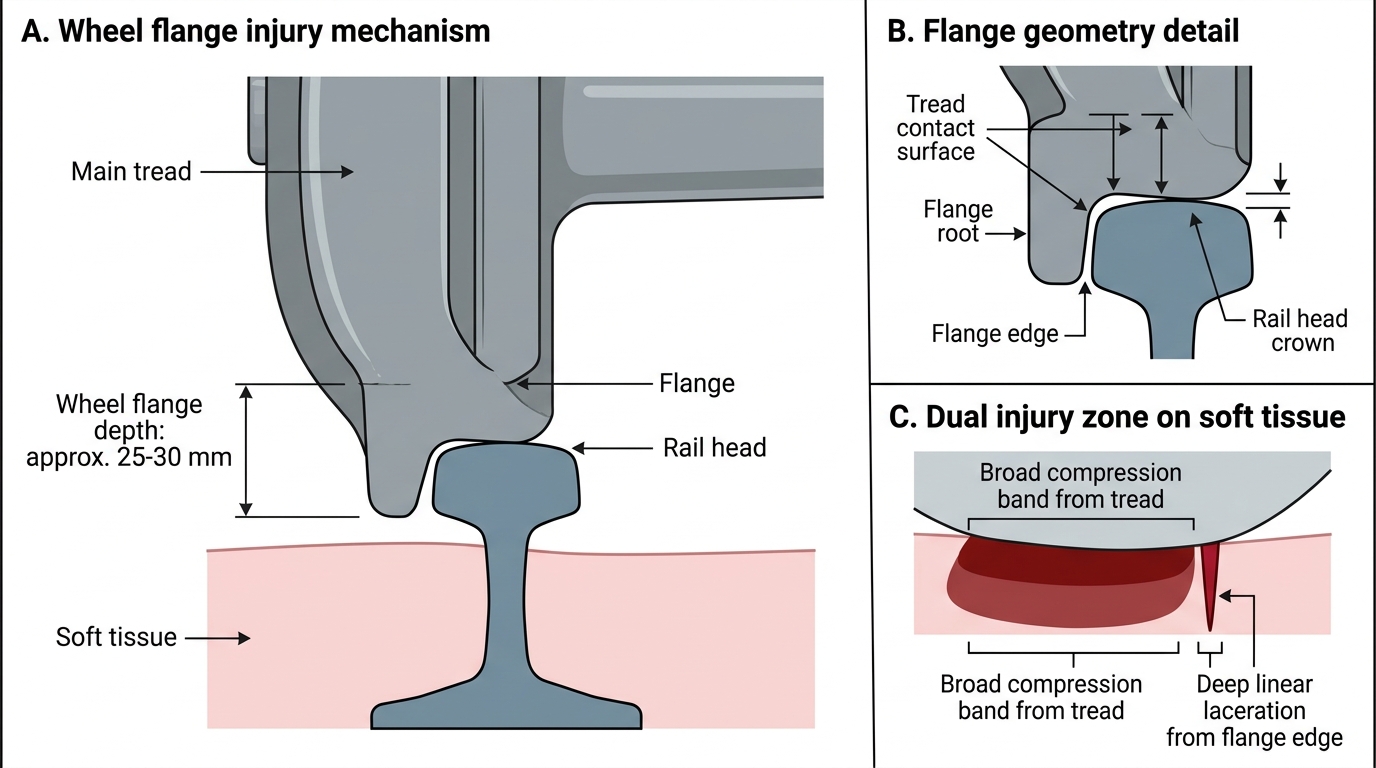
Railway Wheel Flange Injury Pattern
CLINICAL PEARL
The bumper fracture height is a vehicle identifier — but only if the victim was standing upright. A common forensic error is measuring the bumper fracture height on a victim who was crouching, kneeling, or partially supine at the time of impact — for example, a motorcyclist who was already falling off their bike. In such cases, the fracture height does not correspond to the bumper height at all. Before using the bumper fracture height to identify the vehicle, always consider: was the victim standing upright? Is the fracture height physiologically consistent with the reported impact height? Are there scene indicators (bicycle, motorcycle, kerb) that suggest the victim's legs were in an unusual position? Report the bumper fracture height as 'consistent with a vehicle whose bumper is approximately X cm in height, assuming the victim was standing upright at the time of impact' — the qualifying clause protects your opinion from challenge.
Crush Syndrome: Mechanism and Medicolegal Significance
Crush syndrome (traumatic rhabdomyolysis) is a systemic consequence of prolonged mechanical compression of large muscle masses — most commonly seen in earthquake victims trapped under rubble, industrial accidents, and victims of vehicular entrapment. It is distinct from crush fractures and from simple acute compression injuries: crush syndrome refers to the metabolic and renal consequences of massive rhabdomyolysis that follows release of the compression.
The pathophysiology begins with prolonged muscle compression — typically lasting 4–6 hours or longer, though shorter periods can cause syndrome in large muscle groups. Sustained compression causes muscle cell ischaemia and mechanical disruption, destroying the sarcolemmal cell membrane. On release of compression (rescue of the victim), the damaged muscle cells release their intracellular contents into the circulation: myoglobin (a large oxygen-carrying protein), creatine kinase (CK), potassium, and uric acid.
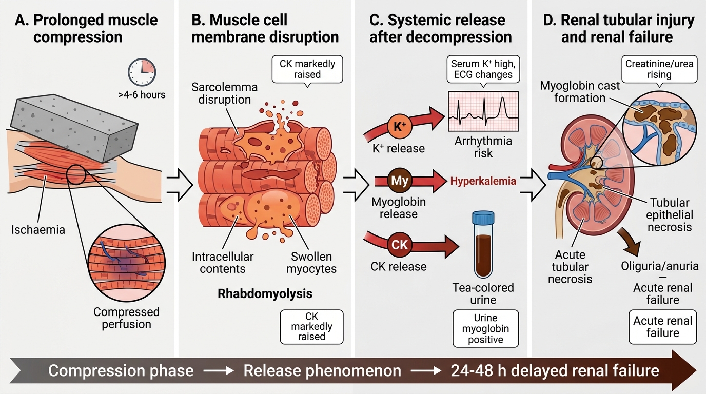
Crush Syndrome Pathophysiology
The systemic consequences are dual:
Hyperkalemia from massive intracellular potassium release can cause fatal cardiac arrhythmias within minutes of release — the 'lethal release phenomenon.' This is why crushing victims can die suddenly at the moment of extraction, even when they have been conscious and communicating while trapped.
Myoglobinuria — myoglobin filtered by the glomerulus at concentrations exceeding renal tubular capacity precipitates in the tubules, causing cast formation and acute tubular necrosis (ATN). The urine turns a characteristic tea-colored or dark brown (myoglobin). ATN leads to acute renal failure with oliguria progressing to anuria. The classic delayed presentation of crush syndrome is the victim who appears well immediately after rescue but develops oliguria, uraemia, and peripheral oedema 24–48 hours later.
Forensic significance of crush syndrome: (1) in disaster investigations, the forensic physician must determine whether a victim died from the primary crush injuries (structural failure, direct organ damage) or from the systemic consequences (ATN, arrhythmia). This affects the estimate of survival time and may be relevant to insurance and liability claims. (2) In forensic autopsies of crush victims, histological examination of the kidneys (tubular casts containing myoglobin/haemoglobin, ATN morphology) and measurement of ante-mortem CK and renal function tests (if available from the clinical record) confirm the diagnosis. (3) In disaster victim identification, crush syndrome is a marker that the victim was alive and conscious for a significant period after entrapment — relevant in calculating suffering and liability.
Scene Examination and Clinical Documentation of Vehicular Injuries
The forensic physician's role in vehicular injury cases extends from the post-mortem room to the scene — scene examination provides context that cannot be recovered later and may make the difference between a sustainable medico-legal opinion and one that unravels under cross-examination.
At the scene, the physician (or investigator) should record: the road type (rural/urban, highway/local road); road conditions (wet, dry, gravel, pot-holed); lighting conditions; the position of the body relative to the road edge and any vehicle skid marks; the position of any scattered personal effects, shoes, or glasses (indicating the point of initial impact); and vehicle damage. The height and profile of vehicle damage (dented bumper, cracked windscreen, blood or hair on the bonnet) correlates directly with the injury levels documented at post-mortem. Where the police have photographs of the scene and the suspect vehicle, these should be requested and reviewed alongside the post-mortem findings.
In the post-mortem examination of a vehicular death, document each injury group systematically:
- Impact surface abrasions: measure their orientation relative to the body axis (horizontal abrasions suggest the vehicle struck the body laterally; diagonal abrasions suggest an angled impact).
- Bumper fracture: measure height from heel to fracture site (if the victim was barefoot) or to the bottom of footwear sole and then to fracture. Record the fracture pattern (transverse = bumper; oblique/spiral = rotational force component).
- Ground-contact injuries: typically on the dependent surface — assess for skull fractures from secondary ground impact, road abrasions (direction indicates direction of body movement after impact).
- Tyre tread patterns: photograph with scale bar; attempt tread pattern identification.
- Clothing examination: note the direction of fibre displacement at the entry wound of the primary impact (fibres pushed inward at the impact side).
For fall from height cases, the scene examination includes: the height of the fall point (confirmed with building or construction plans if possible), any intermediate structures, the condition of the landing surface, and whether there are any signs of a struggle at the top of the fall point (scuff marks, fabric caught, blood at the edge).
SELF-CHECK
A construction worker is extracted from rubble 6 hours after a building collapse. He is conscious and communicating on rescue. His urine is dark brown. Three hours later he becomes anuric and develops ECG changes (peaked T-waves, widened QRS). What is the diagnosis and what is the most immediately life-threatening laboratory abnormality?
A. Acute haemorrhage from internal injuries; the ECG changes reflect cardiac contusion from chest crush
B. Crush syndrome with rhabdomyolysis; the most immediately life-threatening abnormality is hyperkalemia from massive intracellular potassium release
C. Renal contusion from blunt abdominal trauma; the dark urine is from haematuria, and ECG changes are incidental
D. Liver failure from hepatic crush injury; the dark urine is bilirubin and the ECG changes reflect hepatic encephalopathy
Reveal Answer
Answer: B. Crush syndrome with rhabdomyolysis; the most immediately life-threatening abnormality is hyperkalemia from massive intracellular potassium release
This is the classic presentation of crush syndrome: prolonged entrapment (6 hours) followed by myoglobinuria (dark brown urine from myoglobin), progressive renal failure (anuria), and hyperkalemia-induced cardiac changes (peaked T-waves and widened QRS are classic ECG signs of hyperkalaemia). Hyperkalemia from massive intracellular potassium release is the most immediately life-threatening complication — it can cause ventricular fibrillation and sudden cardiac death. Myoglobin-mediated ATN causes the delayed renal failure.