Page 12 of 19
PA30.{3,5} | Phyllodes Tumor & Breast Morphology — SDL Guide (Part 3)
Microscopic Morphology Part 4 — Paget Disease, Inflammatory Carcinoma, and Special IHC
Paget disease of the nipple (microscopy):
Large malignant cells (Paget cells) with abundant pale vacuolated cytoplasm and large nuclei scattered individually within the nipple epidermis — they do NOT form glands, they sit as isolated cells in the squamous epithelium. This creates the clinical picture of nipple eczema that fails to respond to steroid creams.
Mechanism: malignant cells from an underlying DCIS or invasive carcinoma migrate up through the ductal system and invade the nipple epidermis via a pagetoid spread mechanism (LCIS can do this too — 'pagetoid spread' is named after this disease).
Key IHC: Paget cells are CK7+, HER2+, mucin+ (CEA positive). Distinguishes from Toker cells (normal clear cells in nipple epidermis, CK7+, HER2−, mucin−) and from melanoma (Melan-A/S100 positive, CK7−).
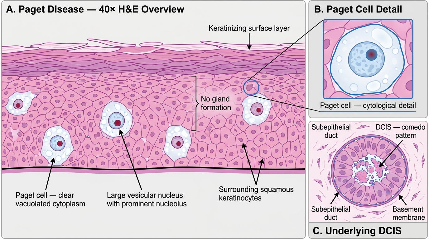
Paget Disease of the Nipple — High-Power Histology with Underlying DCIS
Inflammatory carcinoma (microscopy):
There is no classic microscopic pattern of the underlying tumor — it is most commonly high-grade invasive ductal NST. The diagnostic microscopic finding is dermal lymphatic invasion — clusters of tumor cells within dilated lymphatic channels in the dermis. This is confirmed on a skin punch biopsy. Note: the 'inflammation' of the name is a misnomer — there is NO true inflammatory infiltrate in the dermis (the red, warm, edematous appearance is lymphedema from obstructed lymphatics).
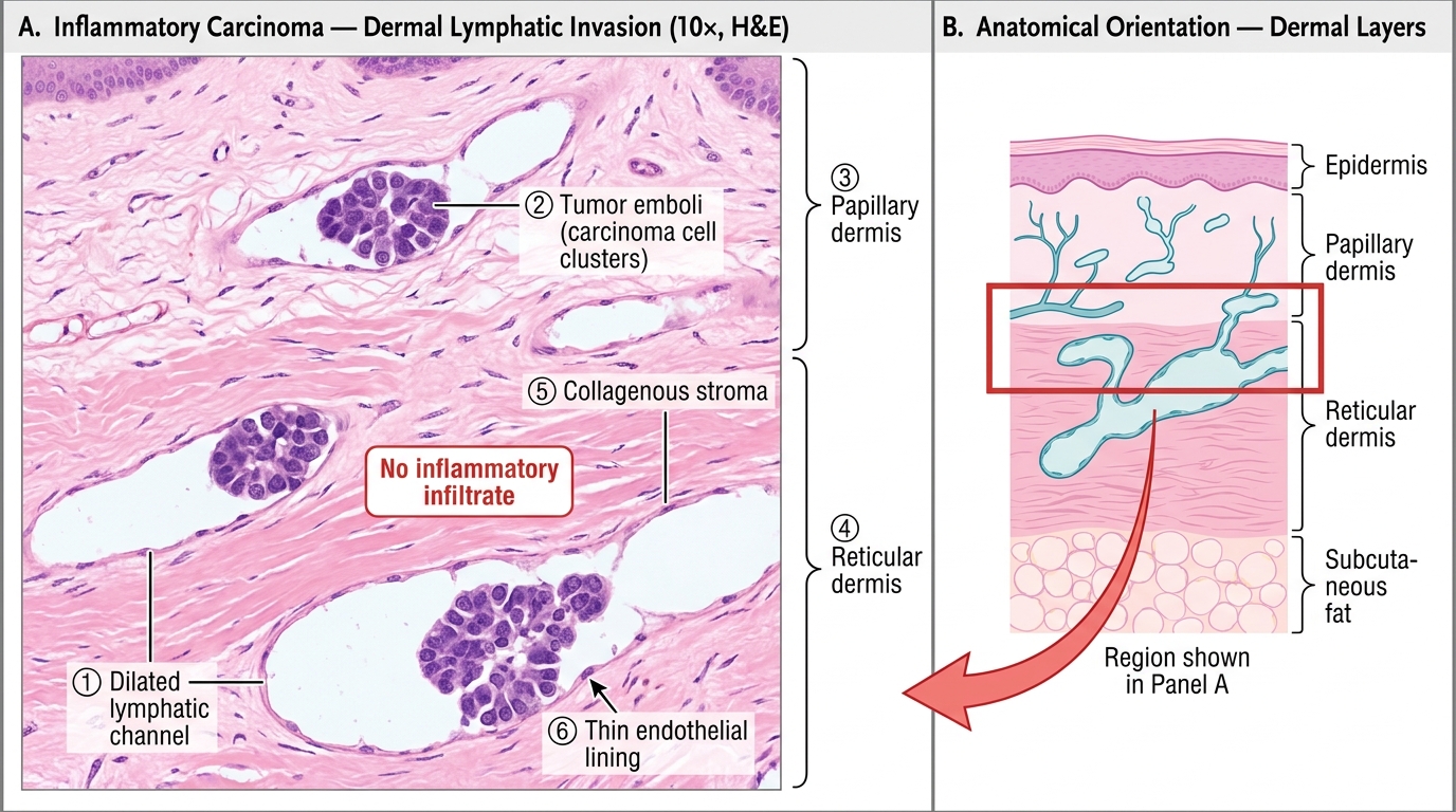
Inflammatory Carcinoma: Dermal Lymphatic Invasion with Tumor Emboli (10×)
Nottingham Histological Grade (Bloom-Richardson-Elston Grade):
All invasive breast carcinomas should be graded using three parameters, each scored 1–3:
1. Tubule/gland formation (> 75% = 1; 10–75% = 2; < 10% = 3)
2. Nuclear pleomorphism (small uniform = 1; moderate variation = 2; marked variation = 3)
3. Mitotic count (per 10 HPF, calibrated to field area — thresholds vary by microscope)
Total score → Grade:
- 3–5 = Grade 1 (well differentiated) — best prognosis
- 6–7 = Grade 2 (moderately differentiated)
- 8–9 = Grade 3 (poorly differentiated) — worst prognosis
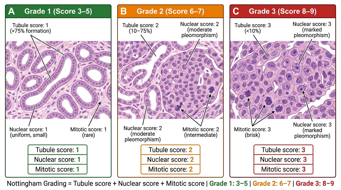
Nottingham Histological Grading of Invasive Breast Carcinoma — Grade 1, 2, and 3 Comparison
SELF-CHECK
A microscopic section from an invasive breast carcinoma shows tumor cells streaming in single-file lines through the fibrous stroma with a targetoid pattern around normal ducts. E-cadherin immunostain is negative. Which type of invasive carcinoma is this?
A. Invasive ductal carcinoma NST
B. Tubular carcinoma
C. Invasive lobular carcinoma
D. Mucinous carcinoma
Reveal Answer
Answer: C. Invasive lobular carcinoma
The Indian-file (single-file) pattern, discohesive cells streaming through stroma in targetoid arrangement around normal structures, and NEGATIVE E-cadherin are the classic features of invasive lobular carcinoma (ILC). E-cadherin loss is the defining molecular event — ILC lacks E-cadherin, making the cells discohesive. Ductal NST forms nests and sheets. Tubular forms angulated glands. Mucinous shows cells in mucin pools.
Master Recognition Table: Practical Exam Pattern Summary
Use this table as a rapid-revision reference. In the practical exam, your answer should cover: lesion name → key gross feature → key microscopic feature → one distinguishing point.
| Lesion | Gross | Microscopy — Key Feature | Distinguishing Point |
|---|---|---|---|
| Fibroadenoma | Firm, rubbery, white, encapsulated, lobulated | Intracanalicular (slit-like ducts) + pericanalicular (round ducts); hypocellular myxoid stroma | Hypocellular stroma vs phyllodes (hypercellular) |
| Phyllodes tumor (benign) | Large, firm, leaf-like cross-section | Leaf-like fronds; mildly hypercellular stroma; pushing margins; < 5 mitoses/10HPF | Same architecture as fibroadenoma but stroma is hypercellular |
| Phyllodes tumor (malignant) | Large, necrotic, haemorrhagic | Marked stromal cellularity, pleomorphism; ≥10 mitoses/10HPF; stromal overgrowth; infiltrative margins | Stromal overgrowth = epithelium replaced by stroma in ≥1 ×4 field |
| Fibrocystic change | Bilateral cysts (Bloodgood blue-domed) + fibrosis | Cysts + apocrine metaplasia + fibrosis ± ductal hyperplasia | Apocrine snouts; ± ADH if nuclear atypia |
| Intraductal papilloma | Small friable intraluminal wart; bloody nipple discharge | Fibrovascular core + bilayer (epithelial + myoepithelial) | Myoepithelial layer present (lost in papillary carcinoma) |
| DCIS low-grade | Often invisible grossly | Cribriform/micropapillary; uniform cells; intact BM; myoepithelial layer present | No necrosis; low nuclear grade |
| DCIS high-grade (comedo) | Toothpaste-like necrosis from ducts | Large pleomorphic cells; central necrosis; microcalcification | Central comedonecrosis + coarse calcification on mammogram |
| LCIS | No mass | Uniform discohesive cells distending acini; no necrosis | E-cadherin negative; bilateral risk marker |
| IDC NST | Gritty, grey-white, stellate, hard | Irregular infiltrating nests/cords; dense desmoplastic stroma | Desmoplasia — the hallmark |
| ILC | Often no mass; diffuse induration | Indian-file (single-file) infiltration; targetoid growth; E-cadherin negative | Single-file = ILC; E-cad negative |
| Mucinous carcinoma | Soft, gelatinous | Tumor cells in extracellular mucin pools | Gelatinous gross; mucin Alcian blue+; good prognosis |
| Medullary carcinoma | Soft, well-circumscribed | Syncytial sheets; dense lymphocytic infiltrate at periphery; pushing margins | BRCA1; lymphocytic infiltrate = good prognosis despite high-grade |
| Tubular carcinoma | Small, stellate | Well-formed angulated tubules, single layer; no myoepithelium | Best prognosis; angulated lumens = tubular |
| Paget disease | Eczematous nipple | Large pale Paget cells in nipple epidermis; CK7+/HER2+ | Nipple involvement; does not spare nipple (unlike eczema) |
| Inflammatory carcinoma | Peau d'orange; no mass | Dermal lymphatic invasion — tumor emboli in dermal lymphatics | Not truly inflammatory; T4d staging |
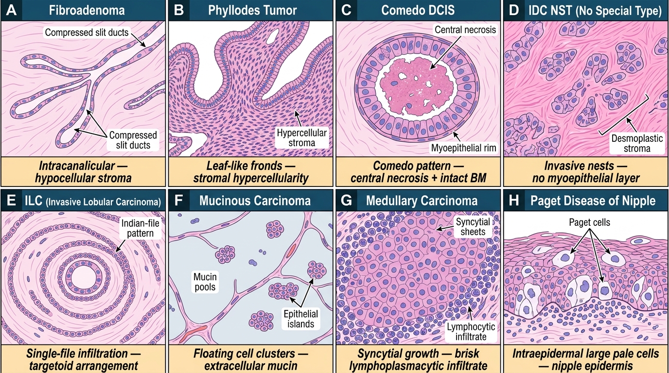
Breast Pathology — Summary Recognition Mosaic (8 Entities)
CLINICAL PEARL
The two most important practical-exam distinctions — say these out loud until they are automatic:
- Fibroadenoma vs Phyllodes: Both are fibroepithelial. The single discriminator at low power is stromal cellularity. Fibroadenoma stroma is pale, sparse, hypocellular. Phyllodes stroma is cellular, crowded, dark. Then check for leaf-like architecture, mitoses, and margins. If you have to say one thing, say: 'The stroma is markedly hypercellular — this is phyllodes, not fibroadenoma.'
- DCIS vs Invasive carcinoma: Stand back and look at the duct contour. In DCIS, the duct is a smooth oval with an outer myoepithelial rim — the tumor fills the inside but the container is intact. In invasive carcinoma, there IS no container — the cells burst out into the stroma in irregular jagged nests without any surrounding myoepithelial layer. These two things cannot look the same once you have trained your eye on them.
SELF-CHECK
Which parameter, when present in phyllodes tumor, most strongly predicts risk of distant metastasis?
A. Mitotic count of 5–9 per 10 HPF
B. Infiltrative tumor margins
C. Stromal overgrowth (stroma replacing epithelium in ≥1 ×4 field)
D. Moderate stromal nuclear atypia
Reveal Answer
Answer: C. Stromal overgrowth (stroma replacing epithelium in ≥1 ×4 field)
Stromal overgrowth — defined as stroma completely replacing the epithelial component in at least one low-power (×4) field — is the single parameter most strongly associated with distant metastasis in phyllodes tumor. It signals that the stroma is behaving as a pure sarcoma. Mitotic count, margin status, and nuclear atypia all contribute to overall grading, but stromal overgrowth is the most ominous individual finding. Its presence automatically places the lesion in the malignant grade.