Page 5 of 20
EN4.35 | Salivary Gland Diseases — SDL Guide (Part 2)
Clinical Assessment: History and Examination of Salivary Gland Disease
A structured clinical approach to salivary gland disease separates the common from the serious and guides investigation decisions efficiently. The clinical encounter is particularly rewarding in salivary gland pathology because a well-taken history often generates a confident differential before any investigation is ordered — the mealtime syndrome is practically diagnostic of a calculus, bilateral sicca symptoms in a middle-aged woman point strongly to Sjögren's syndrome, and rapid growth with facial weakness in a parotid mass demands urgent malignancy workup. Physical examination of the salivary glands requires thorough knowledge of their anatomical positions, their ductal opening sites, and the technique of bimanual palpation — a specific clinical skill tested in ENT practical examinations at the MBBS level. The examination must always include a formal assessment of facial nerve function when the parotid is involved, since facial palsy is the single most important sign of parotid malignancy.
History — key questions:
1. Onset and duration: acute (<2 weeks) suggests inflammation or infection; chronic (months to years) suggests tumour, chronic sialadenitis, or systemic disease.
2. Pain: mealtime swelling and pain that subsides postprandially is pathognomonic of ductal obstruction (salivary calculus). Pain in a long-standing swelling, especially if rapid recent growth, is a red flag for malignant transformation.
3. Relation to meals (mealtime syndrome): specific for sialolithiasis.
4. Dry mouth (xerostomia) and dry eyes (xerophthalmia): suggest Sjögren's syndrome.
5. Facial weakness or numbness: facial palsy in association with a parotid mass is a major red flag for malignancy (facial nerve involvement by tumour). Facial pain/paraesthesia suggests perineural invasion (adenoid cystic carcinoma).
6. Systemic symptoms: fever and malaise suggest acute infection; weight loss and night sweats suggest lymphoma or HIV.
7. Drug history: anticholinergics, antidepressants, diuretics cause xerostomia; phenothiazines and antihypertensives can cause salivary gland swelling (sialadenosis).
Physical examination — principles:
- Inspection: note site (parotid, submandibular, floor of mouth), size, skin changes (erythema — infection; fixed skin — malignancy), and facial symmetry.
- Bimanual palpation: one gloved finger in the floor of the mouth along Wharton's duct, other hand externally over the submandibular triangle — feel for a calculus (hard, palpable along the duct course) and assess gland tenderness and consistency.
- Duct inspection: examine Stensen's duct orifice (opposite upper second molar) and Wharton's duct orifice (sublingual papilla) for redness, discharge, or calculus. Milk the gland from behind forward — clear watery saliva is normal; turbid saliva or pus indicates infection; no secretion indicates obstruction.
- Consistency of the swelling: soft and fluctuant suggests abscess or Warthin's; firm and non-tender suggests pleomorphic adenoma; hard, irregular, and fixed suggests malignancy.
- Facial nerve assessment: test all five branches. Any facial weakness in association with a parotid mass requires urgent FNAC and imaging.
- Neck lymph nodes: cervical lymphadenopathy in association with a salivary gland mass raises concern for malignancy with nodal spread or a systemic cause (lymphoma, sarcoidosis, HIV).
Investigations for Salivary Gland Disorders
Investigation choice depends on the clinical differential but follows a logical progression from cheap and non-invasive to more specialised.
Ultrasound (USS) is the first-line imaging modality for any salivary gland swelling. It is inexpensive, widely available, has no radiation, and can distinguish solid from cystic lesions, identify calculi (posterior acoustic shadowing), assess gland parenchymal changes, and guide FNAC. USS cannot reliably differentiate benign from malignant solid masses — it provides architecture but not histology.
Plain X-ray (dental panoramic — OPG, or lower occlusal view): approximately 80% of submandibular calculi are radio-opaque (calcium phosphate/oxalate) and will be visible on a plain radiograph; approximately 50% of parotid calculi are radio-opaque. A lower occlusal view shows the submandibular duct floor of mouth; the OPG shows the submandibular gland region and Stensen's duct calculi near the parotid. A lateral soft-tissue neck X-ray can show a submandibular duct stone in profile.
Sialography: contrast is injected into Stensen's or Wharton's duct under fluoroscopy — it demonstrates ductal anatomy, strictures, calculi, sialectasis (beaded dilated ducts in chronic parotitis), and parotid lobulation. Contraindicated in acute infection (risks driving infection deeper).
CT neck with contrast: indicated when USS shows a deep or complex mass or when malignancy is suspected. CT demonstrates bone erosion, deep lobe parotid tumours extending through the stylomandibular tunnel, and cervical lymph node involvement. Superior spatial resolution vs MRI for calculi.
MRI with gadolinium: preferred over CT for soft-tissue characterisation of salivary gland tumours and perineural spread (e.g., adenoid cystic carcinoma along the facial nerve). MRI demonstrates extraglandular extension, parapharyngeal involvement, and skull base infiltration better than CT.
Fine-needle aspiration cytology (FNAC): the key pre-operative diagnostic tool for salivary gland masses. Under USS guidance, a 22–25-gauge needle aspirates cells from the lesion. FNAC can distinguish benign tumours (pleomorphic adenoma, Warthin's tumour) from malignant ones (mucoepidermoid, adenoid cystic, carcinoma ex pleomorphic adenoma) with good sensitivity and specificity. Incisional biopsy of parotid masses is contraindicated — it risks tumour seeding and facial nerve injury.
Serological investigations: anti-Ro (SSA) and anti-La (SSB) antibodies for Sjögren's syndrome; ANA, RF; ACE level (sarcoidosis); HIV serology if relevant; FBC (lymphoma workup).
Minor salivary gland biopsy: of labial minor salivary glands (lower lip), considered the gold standard histological confirmation for Sjögren's syndrome — focal lymphocytic sialadenitis with a focus score ≥1.
Diagnosis and Differential Diagnosis of Salivary Gland Swellings
The differential diagnosis of salivary gland swelling requires systematic consideration of the affected gland, the acuity of onset, and the presence or absence of pain, systemic features, and red-flag findings. The framework is built on three initial questions: Which gland is affected? Is the swelling acute or chronic? Is there pain, and if so, is it constant or meal-related? These three questions, combined with the examination findings described in the preceding section, will cluster the patient into one of the major diagnostic categories with considerable accuracy even before investigations return. The significance of correctly reaching a diagnosis here cannot be understated — a delayed diagnosis of parotid malignancy, or a missed submandibular calculus treated as recurrent cellulitis with repeated antibiotics, represents a failure at the bedside that investigations would have easily resolved.
Parotid swellings:
- Acute, painful, unilateral: acute bacterial parotitis (commonest in elderly, post-op), parotid abscess, infected parotid lymph node.
- Acute, painful, bilateral: mumps parotitis (paramyxoviral, unvaccinated), — distinguish from bilateral reactive parotitis in dehydration.
- Chronic, painless, unilateral: pleomorphic adenoma (commonest — slow-growing, firm, lobulated, mobile, no facial palsy), Warthin's tumour (softer, fluctuant, parotid tail, older males), other benign tumours, low-grade malignancy.
- Chronic, painless, bilateral: Sjögren's syndrome, sarcoidosis (Heerfordt syndrome = fever + uveitis + facial palsy + parotid enlargement), sialadenosis (non-inflammatory, non-neoplastic bilateral parotid enlargement — associated with alcoholism, malnutrition, diabetes, liver disease), HIV-associated lymphoepithelial cysts.
- Chronic, progressive, ± pain or facial palsy: malignant parotid tumour — red flags include rapid growth, hard consistency, fixity to skin or deep tissues, regional lymphadenopathy, and facial nerve palsy or paraesthesia.
Submandibular swellings:
- Episodic, mealtime, ± acute infection: submandibular salivary calculus (mealtime syndrome is highly specific).
- Chronic, firm swelling: submandibular tumour (higher malignant rate than parotid) — requires FNAC; lymph node metastasis from oral cavity primary (check lips/tongue/floor of mouth).
- Inflammatory mass: submandibular lymphadenitis, submandibular abscess.
Distinguishing benign from malignant salivary tumour — clinical pointers:
| Feature | Benign | Malignant |
|---|---|---|
| Growth rate | Slow, years | Rapid, months |
| Pain | Absent (usually) | May be present |
| Consistency | Firm, lobulated | Hard, irregular |
| Mobility | Mobile | Fixed (skin or deep) |
| Facial nerve | Normal | Palsy (parotid malignancy) |
| Lymph nodes | Absent | May be enlarged |
⚑ AI image — pending faculty review (auto-QA score 4/10; best of 3 attempts)
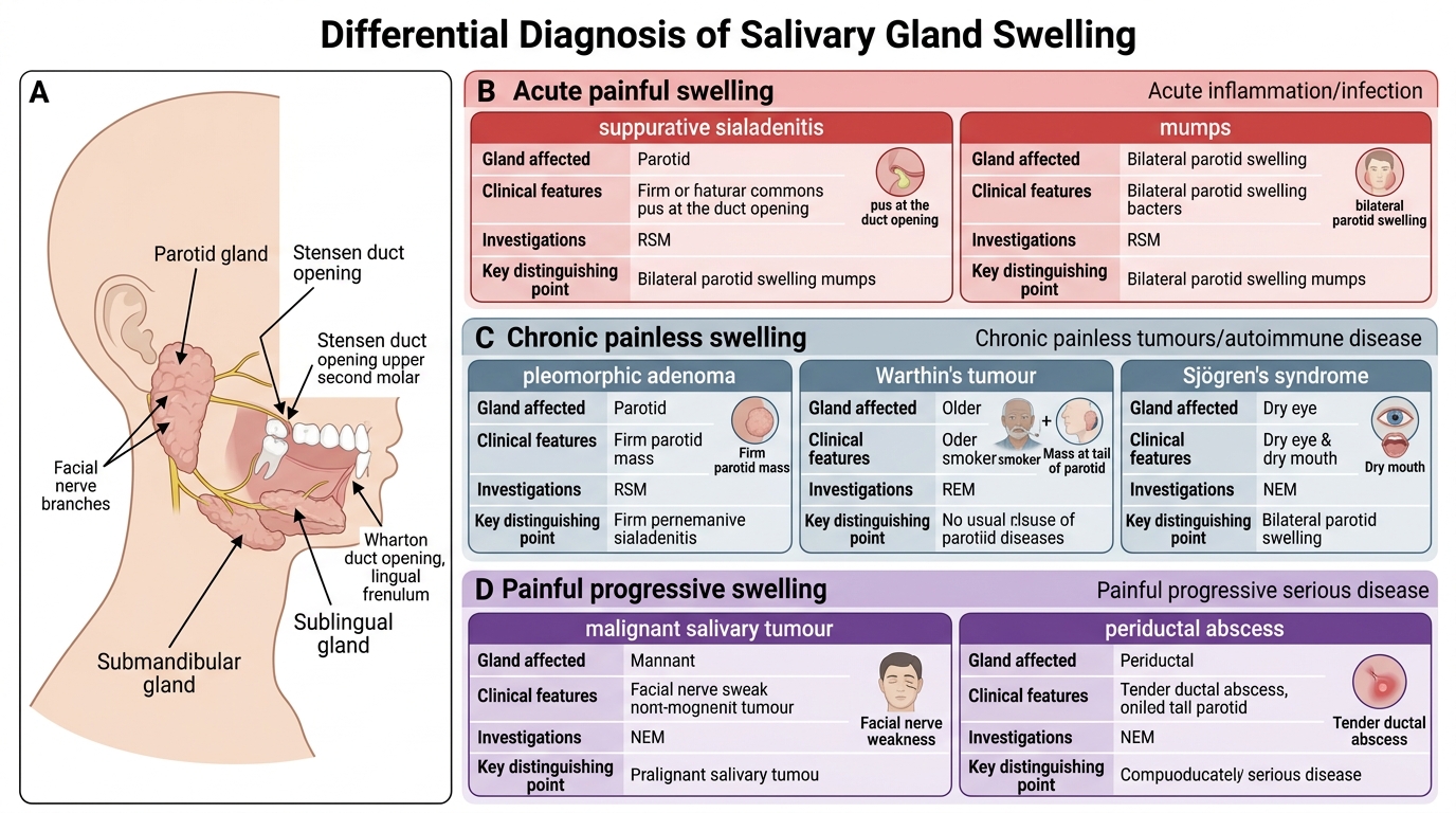
Differential Diagnosis of Salivary Gland Swelling
SELF-CHECK
A 38-year-old woman presents with a 3-year history of bilateral parotid enlargement, dry mouth, and dry eyes. She has been unable to finish a meal without drinking water. Anti-Ro and anti-La antibodies are positive. Lip biopsy shows focal lymphocytic sialadenitis. The diagnosis is:
A. Bilateral parotid sialadenosis due to alcoholic liver disease
B. Primary Sjögren's syndrome
C. Bilateral Warthin's tumour
D. Heerfordt syndrome (uveoparotid fever)
Reveal Answer
Answer: B. Primary Sjögren's syndrome
This is a classic presentation of primary Sjögren's syndrome: bilateral parotid swelling, xerostomia (dry mouth causing need to drink during meals), xerophthalmia (dry eyes), positive anti-Ro/anti-La antibodies, and confirmatory focal lymphocytic sialadenitis on lip biopsy (focus score ≥1). Sialadenosis is painless bilateral parotid enlargement but is non-inflammatory and associated with metabolic causes; it would not produce the sicca complex or positive autoantibodies. Warthin's tumours are unilateral (bilateral in 10%) and do not cause xerostomia or autoantibodies. Heerfordt syndrome is sarcoidosis-related parotid enlargement associated with fever, uveitis, and facial palsy — not the sicca complex or autoantibodies.
CLINICAL PEARL
Two non-negotiable safety rules in parotid surgery: (1) Never perform incisional biopsy on a parotid mass — it risks tumour seeding, facial nerve injury, and Frey's syndrome. FNAC is the correct pre-operative tissue diagnosis. (2) Pleomorphic adenoma must NEVER be enucleated — it has pseudopod projections that extend through the capsule; enucleation leaves tumour behind and leads to multi-focal recurrence that is far harder to operate on safely than the primary. The correct operation is superficial parotidectomy with facial nerve identification and preservation, excising the tumour with a cuff of normal parotid tissue.