Page 7 of 25
PA3.3 | Chronic & Granulomatous Inflammation — SDL Guide (Part 2)
Non-specific (Diffuse) Chronic Inflammation
Non-specific Diffuse Chronic Inflammation
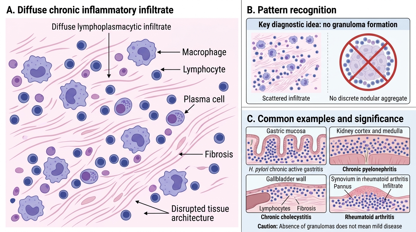
In non-specific chronic inflammation (also called diffuse chronic inflammation), the inflammatory infiltrate of macrophages, lymphocytes, and plasma cells is scattered throughout the tissue without forming organised structures.
Features:
• No granuloma formation
• Accompanying fibrosis often marks the chronic phase
• Tissue architecture is disrupted but no discrete nodular aggregates
Examples:
• Chronic active gastritis (H. pylori) — diffuse lamina propria lymphoplasmacytic infiltrate
• Chronic pyelonephritis — diffuse lymphocytic and plasma cell infiltrate in renal cortex/medulla
• Chronic cholecystitis — lymphocytes and fibrosis in gallbladder wall
• Chronic synovitis in rheumatoid arthritis — diffuse lymphoplasmacytic infiltrate with pannus formation
The absence of granulomas does NOT mean mild disease — chronic non-specific inflammation in rheumatoid arthritis destroys joints; in H. pylori gastritis it leads to gastric adenocarcinoma over decades.
The Granuloma — Definition and Structure
A granuloma is a focal area of granulomatous inflammation characterised by an aggregate of activated macrophages (epithelioid histiocytes), often surrounded by lymphocytes, with or without central necrosis.
Core components:
- Epithelioid histiocytes — the defining cells. Pale, eosinophilic, abundant cytoplasm; vesicular (open-face) nucleus; reduced phagocytic capacity but enhanced secretory function (TNF-α, IL-12).
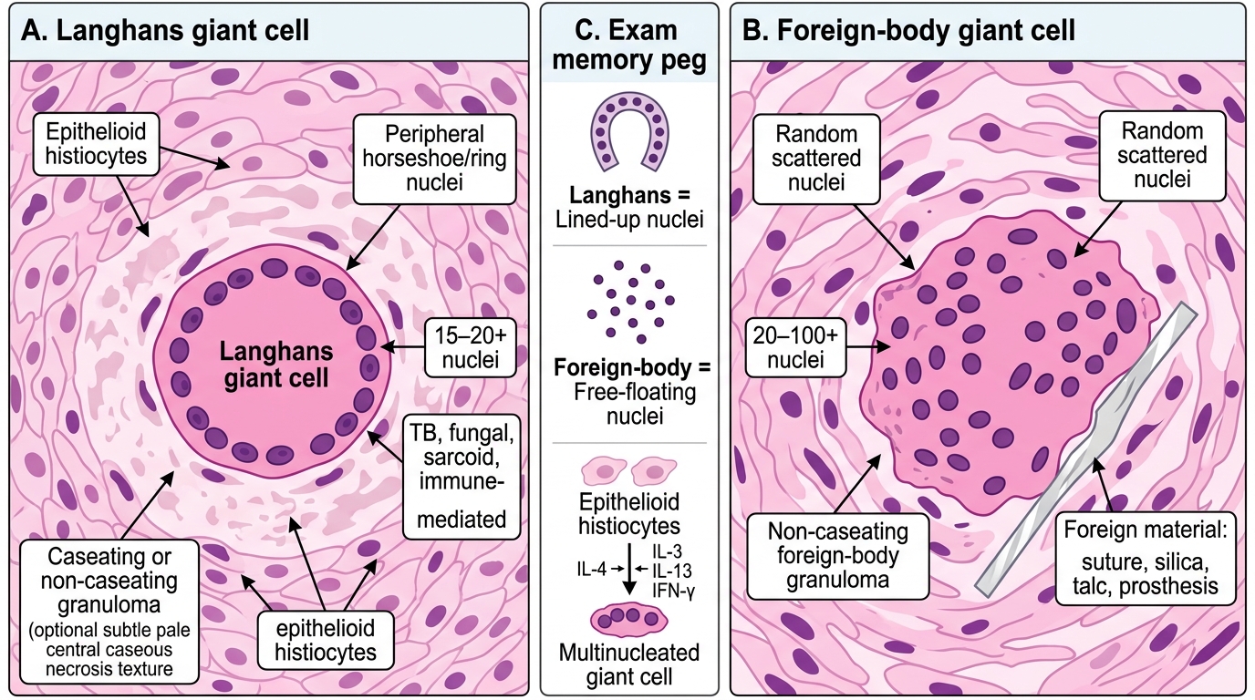
2. Multinucleate giant cells — formed by fusion of epithelioid histiocytes. Two patterns:
• Langhans giant cells: Nuclei arranged in a horseshoe or peripheral ring. Classic for TB and fungal infections.
• Foreign-body giant cells: Nuclei randomly scattered ("jumbled") throughout cytoplasm. Seen around non-digestible foreign material (sutures, talc, silica).
- Lymphocytic cuff — CD4+ Th1 cells surrounding the epithelioid aggregate, maintaining macrophage activation via IFN-γ.
- Central necrosis — present in caseating granulomas (see next section); absent in non-caseating types.
- Fibroblasts and collagen — at the periphery in older or resolved granulomas.

Granuloma: Definition and Structure
Giant Cell Types — Langhans vs Foreign-Body
Recognising the two giant cell types at microscopy is a high-yield, frequently examined skill.
| Feature | Langhans Giant Cell | Foreign-Body Giant Cell |
|---|---|---|
| Nuclear arrangement | Peripheral horseshoe or ring | Randomly scattered (central and peripheral) |
| Size | Large (up to 40–50 µm) | Large, often irregular shape |
| Number of nuclei | 15–20+ | 20–100+ |
| Associated granuloma | Caseating (TB) or non-caseating (fungal, sarcoid) | Non-caseating |
| Clinical context | Infectious or immune-mediated | Foreign material (suture, silica, talc, prosthesis) |
Memory peg: Langhans → Lined-up nuclei (horseshoe). Foreign-body → Free-floating nuclei (random).
Both types form by fusion of epithelioid histiocytes under IL-4, IL-13, and IFN-γ signalling.
Langhans vs Foreign-Body Giant Cells