Page 16 of 32

PA24.{5,7} | Portal Hypertension & Hepatocellular Carcinoma — SDL Guide (Part 3)

Aetiology of Hepatocellular Carcinoma

Infographic showing major causes of hepatocellular carcinoma, including chronic HBV, chronic HCV, aflatoxin B1, and cirrhosis-related metabolic or genetic liver diseases.

Aetiology of Hepatocellular Carcinoma

Panel A: Chronic liver injury, cirrhosis, dysplastic nodules, hepatocellular carcinoma arising from hepatocytes. Panel B: HBV DNA integration, HBx protein, genomic instability, direct oncogenesis, HCV chronic inflammation, fibrosis, cirrhosis-mediated oncogenesis. Panel C: Aflatoxin B1, Aspergillus flavus/parasiticus, stored groundnuts and maize, CYP activation, epoxide, DNA adduct, TP53 codon 249 R249S mutation, HBV synergy. Panel D: Alcoholic cirrhosis, NASH/NAFLD, haemochromatosis, alpha-1 antitrypsin deficiency, primary biliary cholangitis, Wilson disease, cryptogenic cirrhosis as burnt-out NASH.

Hepatocellular carcinoma (HCC) is the most common primary liver malignancy, arising from hepatocytes. It almost always arises on a background of chronic liver injury.

Major aetiological factors:

  1. Chronic HBV infection — globally most important cause; integrates viral DNA (HBx protein) into the hepatocyte genome, causing genomic instability and direct oncogenesis even before cirrhosis. In India, HBV accounts for ~50% of HCC cases.
  1. Chronic HCV infection — predominantly through cirrhosis-mediated oncogenesis (unlike HBV, HCV does not integrate into DNA); ~25% of HCC in India.
  1. Aflatoxin B1 — produced by Aspergillus flavus/parasiticus contaminating stored groundnuts and grain (maize, sorghum) in humid conditions. Critically relevant in India, sub-Saharan Africa, and South-East Asia. Aflatoxin B1 is activated by CYP enzymes to an epoxide that causes a specific TP53 codon-249 mutation (R249S). Synergistic with HBV — the combination raises risk ~60-fold.
  1. Cirrhosis of any cause — alcoholic cirrhosis, NASH-related cirrhosis (rising rapidly with the obesity epidemic), haemochromatosis (10–30% lifetime risk in homozygotes), α1-antitrypsin deficiency, primary biliary cholangitis, Wilson's disease.
  1. Alcohol — acts primarily through cirrhosis; no direct carcinogenic effect equivalent to aflatoxin.
  1. NASH/NAFLD — now a major driver in non-drinkers; 'cryptogenic' cirrhosis progressing to HCC often represents burnt-out NASH.

Pathogenesis — From Cirrhosis to Carcinoma

Sequential medical diagram showing chronic liver injury progressing through regenerative and dysplastic nodules to hepatocellular carcinoma, with key molecular events and HBV-specific oncogenesis highlighted.

Pathogenesis of Hepatocellular Carcinoma

Panel A: Normal liver; chronic injury from HBV/HCV, aflatoxin, alcohol; hepatocyte death; regenerative nodule; low-grade dysplastic nodule; high-grade dysplastic nodule; hepatocellular carcinoma; fibrous septa; malignant transformation.. Panel B: Telomere shortening; chromosomal instability; TP53 mutation, R249S from aflatoxin; loss of apoptosis checkpoint; TERT promoter mutation; telomerase reactivation; Wnt/beta-catenin activation; CTNNB1 mutation; VEGF angiogenesis.. Panel C: HBV integration; HBx protein; TP53 inhibition; growth-factor gene activation; HCC without cirrhosis; hypervascular HCC nodule; arterial vessels; arterial enhancement on CT/MRI..

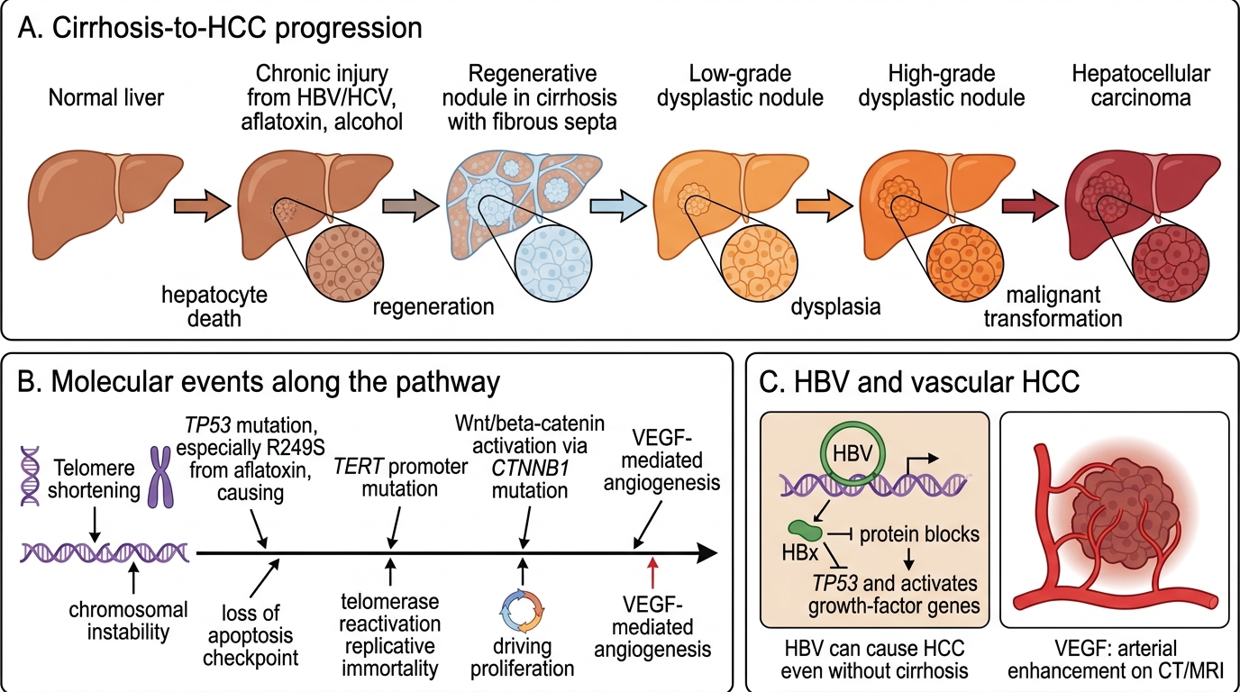
The dominant pathway is: chronic injury → hepatocyte deathregenerative nodulesdysplastic nodulesHCC.

A six-stage schematic shows progression from normal liver through chronic injury, regenerative and dysplastic nodules, to hepatocellular carcinoma with key molecular events labeled.

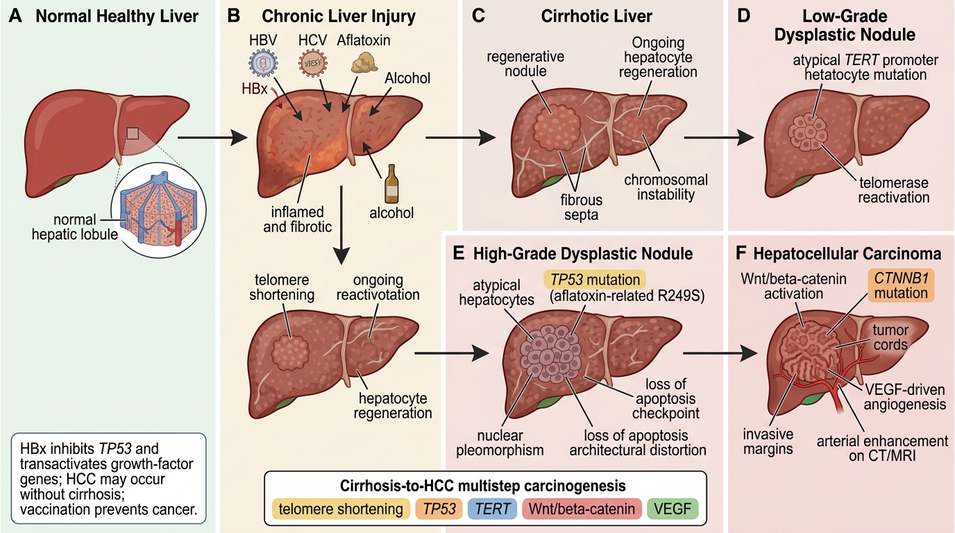
Cirrhosis-to-Hepatocellular Carcinoma Progression

Panel A: Normal liver; smooth hepatic surface; normal hepatic lobule inset; normal hepatocytes. Panel B: Chronic injury; HBV, HCV, aflatoxin, alcohol; inflammation; early fibrosis; HBx protein callout. Panel C: Regenerative nodule; cirrhosis; fibrous septa; regenerating hepatocytes; telomere shortening; chromosomal instability. Panel D: Low-grade dysplastic nodule; mild hepatocyte atypia; early TERT promoter mutation; telomerase reactivation. Panel E: High-grade dysplastic nodule; marked atypia; architectural distortion; TP53 mutation; aflatoxin-associated R249S; loss of apoptosis checkpoint. Panel F: Hepatocellular carcinoma; invasive tumor nodule; Wnt/beta-catenin activation; CTNNB1 mutation; VEGF angiogenesis; arterial enhancement on CT/MRI.

Key molecular events:
Telomere shortening in regenerating hepatocytes → chromosomal instability.
TP53 mutation (especially R249S from aflatoxin) — loss of apoptosis checkpoint.
TERT promoter mutation — the commonest somatic mutation in HCC (~60%); reactivates telomerase, conferring replicative immortality.
Wnt/β-catenin activation (~30%) — CTNNB1 mutations drive proliferation.
VEGF and angiogenic signals — HCC is highly vascular (exploited in imaging: arterial enhancement on CT/MRI).

In HBV-related HCC, the HBx protein transactivates growth-factor genes and inhibits TP53 directly, enabling oncogenesis even without cirrhosis — explaining why HBV vaccination in childhood is a genuine cancer prevention strategy.

Gross Morphology of HCC

Comparison diagram showing unifocal, multifocal, and diffuse gross patterns of hepatocellular carcinoma with a schematic of portal vein and hepatic venous invasion.

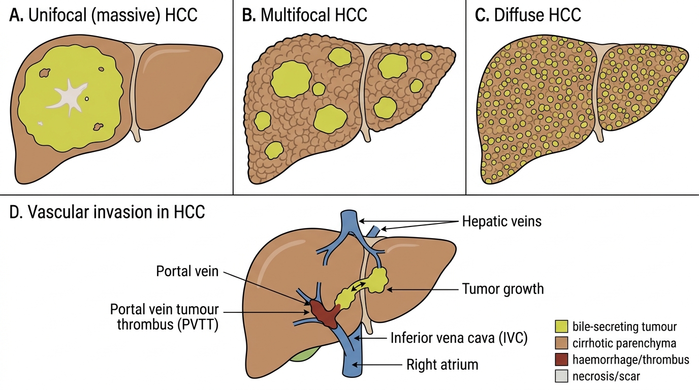
Gross Morphology and Vascular Invasion of HCC

Panel A: Unifocal massive HCC; right lobe; single large yellow-green tumour mass; central scar; necrosis; surrounding liver parenchyma.. Panel B: Multifocal HCC; multiple discrete tumour nodules; varying nodule sizes; cirrhotic background; right lobe; left lobe.. Panel C: Diffuse HCC; innumerable small tumour nodules; cirrhosis-mimicking nodules; diffuse liver involvement.. Panel D: Portal vein tumour thrombus (PVTT); portal vein; hepatic veins; inferior vena cava; right atrium; tumour extension pathway; haemorrhagic thrombus..

Three gross patterns are recognised:

1. Unifocal (massive) — single large tumour, often replacing an entire lobe; commonest in younger patients with HBV without established cirrhosis. May show a central scar or areas of necrosis.

2. Multifocal — multiple discrete nodules of varying size scattered throughout the liver; typically in a cirrhotic background, arising from independent foci of transformation ('multicentric origin') or intrahepatic satellite metastases.

3. Diffuse — innumerable small tumour nodules permeating the entire liver, mimicking cirrhotic nodules macroscopically; difficult to diagnose radiologically.

Colour: Yellow-green (bile-secreting), green-brown, or white with haemorrhagic foci.

Three side-by-side liver gross pathology panels compare unifocal, multifocal, and diffuse hepatocellular carcinoma patterns.

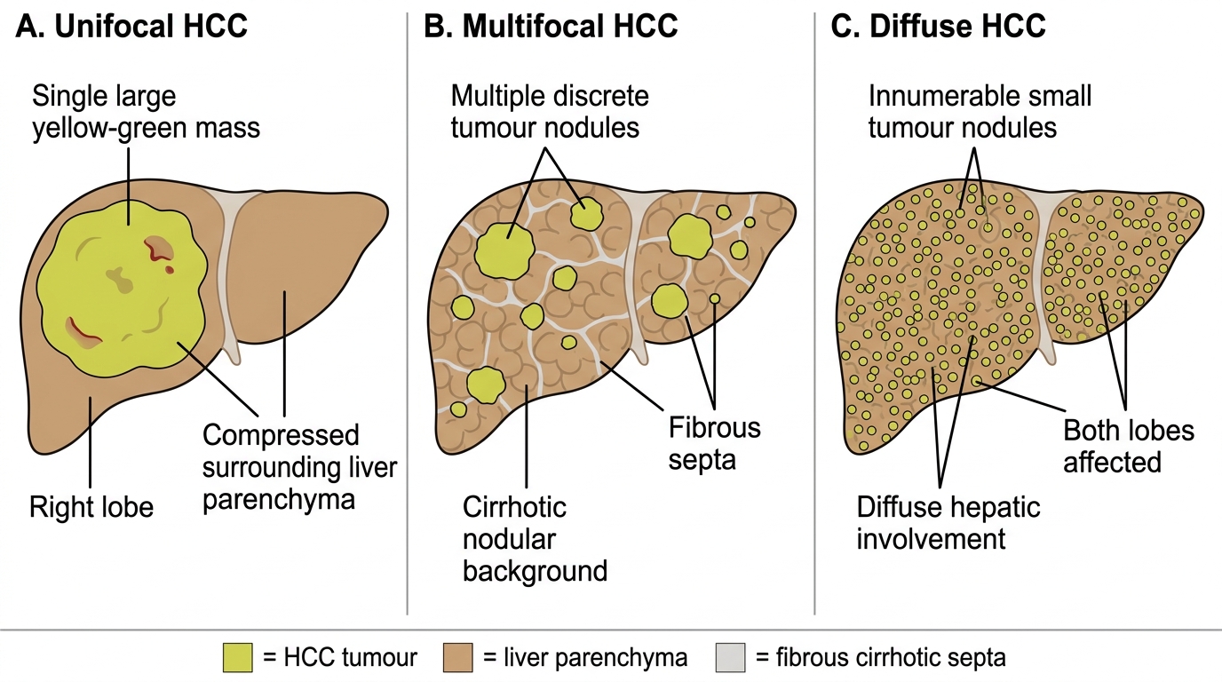
Gross Types of Hepatocellular Carcinoma

Panel A: Unifocal HCC showing a single large yellow-green mass in the right lobe, compressed surrounding liver parenchyma, and necrotic or hemorrhagic cut-surface areas.. Panel B: Multifocal HCC showing multiple discrete yellow-green tumour nodules on a cirrhotic nodular background with fibrous septa.. Panel C: Diffuse HCC showing innumerable small yellow-green tumour nodules distributed throughout both liver lobes..

Vascular invasion: HCC has a striking propensity to invade vascular channels — particularly the portal vein (portal vein tumour thrombus, PVTT) and hepatic veins. PVTT exacerbates portal hypertension, worsens ascites, and carries a dire prognosis. Tumour may extend into the IVC and right atrium.